
A Path to Identifying a Bacteria
- crispincisco
- Apr 20
- 2 min read
Updated: May 4

Initially, bacteria might seem impossible to tell apart. They are microscopic and can not be seen with the naked eye. So, how do scientists identify and tell them apart? Throughout the years, scientists have explored ways to visualize, identify, and differentiate bacterial species. One of the techniques created and refined over the years to identify bacteria in science (microbiology) is called Gram staining.
Understanding the Gram-staining process

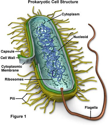
The Gram staining procedure was developed in 1884 by a scientist called Hans Christian Gram. The technique allows scientists to identify bacteria into two large groups, Gram-positive and Gram-negative. The groups are not distinguished by name; they differ structurally, which affects how bacteria behave and respond to antibiotics.
Procedure for Gram-staining

The Gram staining procedure involves staining bacteria using a series of dyes. Before the staining process begins, scientists use a flame (Bunsen burner) to sterilize a loop and place bacteria on a slide, put a drop of water on the bacteria on the slide, and wait a few minutes until it is dry. They then heat-fix the bacterial smear on the slide by running it through the Bunsen burner, before beginning the staining process with the dyes.
First, one or two drops of crystal violet dye are applied to the smear for one minute, then rinsed with water.
Next, the dye is fixed by adding Iodine to the smear for one minute, binding the crystal violet dye to the bacteria on the slide.
Subsequently, a decolorizer (alcohol), often a solvent of ethanol or acetone, is used to remove the dye; it is time sensitive, should last for approximately 10 seconds, and should never be placed directly on the smear, but above it before rinsing with water.
Lastly, safranin (or a fuchsin stain), a counterstain, is used to give a decolorized Gram-negative bacterium a pink color for identification.

Purpose of Gram-Staining.

So, why does this happen? The answer to this question comes down to the bacterial cell wall structure. The stain is applied and binds to the peptidoglycan component of the bacterial cell wall. Gram-positive bacteria have a thick peptidoglycan cell wall that holds on to the crystal violet dye, so they stay purple. Gram-negative bacteria, on the other hand, have a thinner or no peptidoglycan cell wall. When the alcohol wash is applied, the purple dye is washed away, and they pick up the pink dye instead from the fuchsin stain or safranin.
Conclusion
Gram staining is one of the first steps scientists use when studying unknown bacteria. It provides quick information about the bacteria and helps narrow down their identity. Gram staining is an interesting procedure that reveals so much information about bacteria with a simple color change. It turns something invisible into something scientists can categorize, study, and understand. Gram staining is a great example of how small details in science can have big impacts. Through changing the color of bacteria, scientists are able to uncover important information about bacteria that helps drive research, medicine, and our understanding of the microbial world.
Comments