
No More Blind Dates with Your Bacteria!
- as24555
- Nov 6, 2025
- 3 min read
Updated: Nov 10, 2025

Imagine you are a scientist and you have some bacterial growth on a plate. You work with this bacteria and you form what you think is a bond with it because you are a scientist and sometimes scientists are delusional.
So you're working with this bacteria and it behaves a certain way on the plate, but you have no idea what your bacteria looks like individually. You don't know the shape or how big it is or any individual fact about it because it grows in colonies.
Well do I have the procedure for you! This procedure will help you view your bacteria under a microscope and figure out what it looks like! It may even make you more delusional and form a better "bond" with your bacteria because now you will know what it looks like.
This procedure is none other than Gram staining!
Gram staining is a simple procedure that uses stains, decolorizers, and counterstains to let you associate a color - purple or pink - with the type of bacteria you have, as well as see the shape of your bacteria.

The steps are as follows:
Add a drop of water onto a slide
Flame an inoculating loop until it glows red
Let it cool
Pick a colony and smear it onto the water
Wait for the water to evaporate, then heat fix by swiping slide through the flame 3x
Add a few drops of crystal violet
Wait 60s and then wash off with distilled water
Add a few drops of Gram's iodine
Wait 60s then wash off with distilled water
Drip ethanol on the slide until it runs clear, or until 10 seconds has passed
Rinse with distilled water
Add a few drops of safranin
Wait 60s then wash off with distilled water.
Pat dry with bibulous paper then view under a microscope
Now you're ready to view!
Place dry slide onto the microscope stage
Make sure the light is turned on and the magnification is set to 10x lens
Focus the microscope so the bacteria are clearly seen
Then add a drop of immersion oil onto the slide and switch the magnification setting to the 100x lens
Re-focus and identify your bacteria
Move the magnification setting back down to 10x
Wipe off the 100x lens
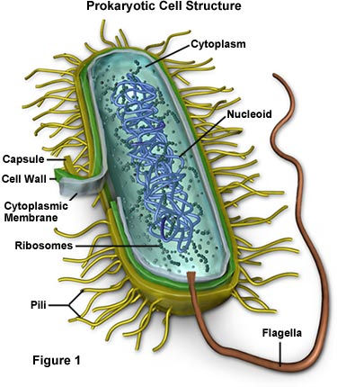
When you view your bacteria under a microscope, you will have the chance to see if the bacteria are gram positive or gram negative, as well as their shape. The difference between gram positive and gram negative lies in the peptidoglycan layers of the cell walls, where gram positive bacteria have thick peptidoglycan layers and appear purple, and gram negative bacteria have thin peptidoglycan layers, and appear pink.

Additionally, you might see different shapes in your sample. These different shapes are the actual shape of your bacteria. If you see little circles, or rods, or spiral-shapes, these are all common shapes of bacteria. Multiple shapes in one sample can show either contamination or show some weird bacteria. Some bacteria are a combination of shapes, and they can make the Gram staining results confusing or inconclusive.
Although Gram staining isn't a perfect technique, as it can give weird results if the steps weren't followed properly, or might show inconclusive results based on the type of bacteria, it is generally a good and quick way of telling what your little bacteria friends look like so you can form deeper attachments to them.
Comments