
We Can See It!
- ra11876
- Apr 2, 2025
- 2 min read
Updated: Apr 21, 2025
Have you ever wondered how scientists distinguish between different types of bacteria under microscopes? One of the most essential techniques in microbiology and in any lab is Gram staining. Gram staining was a method that was developed by Hans Christian Gram in 1884. Although the procedure is simple and fun for most, it is a very powerful procedure that separates bacterium into gram-positive and gram-negative groups based on their cell wall composition.

Why Gram Staining?
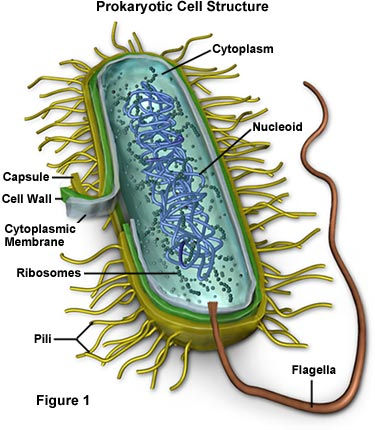
Bacterium is clear and be seen with the naked eye which is where Gram staining comes in! Bacterium have different cell wall structures which determines how they interact with the stains. Gram-positive bacterium have a thick peptidoglycan layer which retains the primary stain and appears purple under the microscope. Gram-negative bacterium have a thin peptidoglycan layer In addition to an outer membrane which causes them to lose the primary stain and take up the counterstain which is pink or red. The distinction between these two colors is critical especially for diagnosing infections and select antibiotics.

How Does It Work?
Gram staining is not a lengthy procedure and can be quite enjoyable. First a drop of water is placed on a clean glass slide. A sterile loop is used to transfer a small amount of bacteria and is mixed with the water and spread into a thin film. Once the slide is air dried the slide is passed through a flame two to three times to kill and fix the bacteria onto the slide. Heat fixation is an important step during staining because it allows the sample to adhere to the slide and kill the bacteria. Four components are added to the slide one by one first crystal violet then grams iodine following ethanol and finally safranin.
Components
Crystal violet: Positively charged which binds to negatively charged components. It is used to initially stain all cells allowing to distinguish between bacterium types based on the cell wall structures.
Grams iodine: Helps fix the crystal violet onto the sample and enhances stain retention in gram-positive cells
Ethanol: Used to wash off the crystal violet-iodine complex. Timing is key, too much of ethanol can cause gram-positives to lose color, too little can cuase gram-negatves to look like gram-positives.
Safranin: Provides contrast to the purple gram-positive cell and makes gram-negatives easier to distinguish after the decolorization step.
Once each stain is added to the sample, individually, It stays on the slide for about 1 minute and then rinsed with distilled water. It is important to know that ethanol is added drop by drop and only for 5 to 10 seconds until the runoff is clear because if ethanol is left on for too long it can decolorize gram-positive cells which can lead to inaccurate results. After this procedure the slide is ready to be placed on a microscope and observe the bacteria sample.
This technique is used in labs by professionals and students. It is a great way to discover and visualize bacterium. Without Mr. Gram the identification of the bacterium we know today would not be possible. Thank you Mr. Gram!
Comments