
Bacteria in Color: What is Gram Staining?
- dhp1016
- Apr 15
- 2 min read
Updated: Apr 20
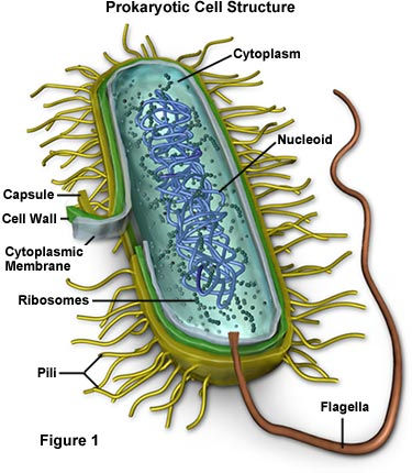
When working with bacteria, it can be hard to visualize them even under a microscope since they are clear but Gram staining allows us to add color and visualize bacteria under a microscope. Gram staining is a very important technique to know and learn when working with bacteria. Gram staining separates bacteria into two major groups, gram-positive and gram-negative, which tells us about the cell wall structure of the bacteria. This is due to the presence of thick protective layers of peptidoglycan in the cell walls of gram-positive bacteria, which gram-negative bacteria lack. This can help us determine how bacteria might respond to different antibiotics and environment.

Steps of Gram Staining:
To perform Gram staining, first you need to put a small number of bacterial cells on a microscope slide and heat fix it then there are 4 steps involved in Gram staining.
First, the slide is flooded with crystal violet that stains all cells, after a minute you wash the slide with distilled water.
Next, Iodine is added that binds to crystal violet and helps it stick to the bacterial cells, which is rinsed with distilled water again after another minute.
Then, the most important step decolorization, where ethanol is added to remove the crystal violet from cell walls of gram-negative bacteria, until the purple stops flowing from the slide.
Finally, a counterstain called safranin that stains everything again, but it is a lighter dye than crystal violet.

So what happens after that?
After Gram staining, you get to see the bacterial cells under a microscope. Using a microscope, we can visualize the results of gram staining which can tell us many things about the bacteria. For example, shape, gram status, and pure of mixed culture. We can tell the shape of the bacteria we are working with (coccus, bacillus, spirochete). Whether it is gram-positive (Purple), gram-negative (Pink), or a mix of both. If there is a mix we know that our culture is not pure but if there isn't a mix we know we isolated a single colony.
Overall, Gram staining is a simple but extremely useful technique in microbiology that can help scientists quickly identify and classify bacteria based on their cell wall structure. By following just four simple steps, we can distinguish between gram-positive and gram-negative bacteria. The results not only tell us about the morphology, but it also tells us whether the sample contains a single species or multiple species of bacteria. Hence, Gram staining is an important first step in studying unknown bacteria and understanding how they may behave under different conditions.

Comments