
Decoding Microbial Morphology: The Steps of Gram Staining
- lad1078
- Apr 1, 2024
- 2 min read
Updated: Apr 8, 2024

Gram staining in microbiology is a fundamental technique that allows for visualization and differentiation of bacteria under the microscope. Through this, we are able to classify bacteria as either gram-positive or gram-negative by the color they appear and place them into categories based on morphology. Join me as I dive into the steps of Gram staining.
Preparation: Prepare the Bacteria Smear

To begin the Gram staining procedure a smear with the desired bacteria must be prepared aseptically and heat fixed before staining. This is done by placing a drop of water on a dry slide with the bacteria and mixing them. Following this, once the slide has completely dried, it can be passed through a flame three times. After the smear has been properly prepared, Gram staining can now begin.

Step 1: Crystal Violet
The first step to a gram stain is to stain the smear with crystal violet which is a primary stain to give all the cells a color. During this step all cells will appear either purple or blue. The crystal violet is to remain on the smear for approximately 1 minute and then rinsed off with distilled water.
Step 2: Gram's Iodine
The second step is to use Gram’s iodine which is a mordant to stabilize stains or dyes by making the dye less soluble as it adheres to the cell walls. During this step all cells will remain either purple or blue. The Gram’s Iodine is to remain on the smear for approximately 1 minute and then rinsed off with distilled water.
Step 3: Decolorize

The third step is to decolorize the gram-negative cell walls using an alcohol such as ethanol or an acetone/ethanol solution. During this step the gram-positive cells will remain purple or blue while the gram-negative cells will become colorless. The alcohol is to run down the smear for approximately 5-10 seconds and no more than that and then rinsed off with distilled water.
Step 4: Safranin

The fourth and final step of the gram stain is to stain the smear with safranin which is a counterstain that highlights the gram-negative cells. During this step the gram-positive cells will remain purple or blue and the gram-negative cells will become pink or red. The safranin is to remain on the smear for approximately 1 minute and then rinsed off with distilled water.
All done!
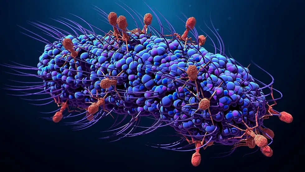
Now you can look at your slide under the microscope where you may see bacteria like the one's below.


Gram staining is a valuable yet simple technique is microbiology giving insight into bacterial classification and morphology. By following these steps, researchers and clinicians can further explore the vast world of microbiology.
Comments