
Introduction to Gram Staining
- hmw1053
- Mar 28, 2024
- 3 min read
Updated: Apr 3, 2024
Disclaimer
This blog posts contains image content that was generated by artificial intelligence. It may contain errors or inaccuracies, and should not be relied upon as a substitute for professional advice.This image content is for entertainment purposes only. It should not be used for any other purpose, such as making financial decisions or providing medical advice.

Gram staining is a valuable diagnostic tool used in clinical and research settings. Although newer molecular techniques have been developed , the gram stain is still a widely used method for the identification of unknown bacteria. It is often one of the first tests conducted on an unknown species in the laboratory, and in some cases it can provide presumptive identification of the organism. For many medical professionals a gram stain of a clinical specimen can be used to determine the appropriate treatment for a bacterial infection.

In 1884 , A Danish doctor named Hans Christian Gram starting experimenting on ways to develop a staining technique that would differentiate bacterial cells from eukaryotic nuclei in diseased lung tissue. Through his experiments he discovered that certain stains were retained by some types of bacterial cells but removed from others during the staining process. He published this work which then served as the most important early staining technique in bacteriology known as the gram stain.

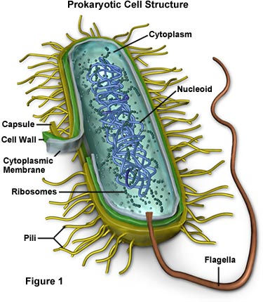
In order for us to fully understand the basic workings of the Gram staining procedure we need to have some knowledge about bacteria. Bacteria can be gram-positive or gram-negative depending on how they will respond to the procedure of gram staining. The bacterial cell has a cell wall, and it will get strength from peptidoglycan and other substances like murein and mucopeptide in this cell wall. This high –strength cell wall is part of the gram staining process.

Cells will be stained with crystal violet and or iodine and then washed with
acetone or alcohol. Due to this procedure the gram-negative bacteria will be
de-colorized, and the gram-positive cells will generally not be. We can de-colorize gram-
positive cells if we use acetone or alcohol then remove the cell wall allowing it to
de-colorized but this step must happen after the staining step and before the washing step. The gram-negative bacteria cell wall has a special function allowing it to be UN-selectively permeable. The outer membrane stops the passage of molecules too large.The staining process will kill the cell due to the stains being composed mainly of salt. However most of the time the bacteria we are working with in our lab is already dead. A classic stain will be colored with a colored cation and a colorless anion.
Fun Fact
Few pathogenic bacteria belong to the gram-positive group and most pathogens are gram-negative.

Because the bacteria are rich with nucleic acids with negative charges (phosphate groups) it can combine with the positively charged dyes.The Gram Stain procedure is considered differential stain because the reactions take advantage of the cells' structure having a dissimilar staining reaction. The general procedure for Gram staining is relatively simple. After we have heat fixed the bacteria of interest to a slide we will start with applying the basic dye crystal violet 1 minute and then rinse it off with sterile H20. Then we will obtain iodine solution and apply it thus staining all the bacteria blue at this point in the procedure 1 minute then rinse again. Then we get some alcohol and treat the cell for 5-10 seconds maximum until the rinse water runs clear. The gram-positive cells will keep the crystal blue iodine color and the gram-negative cells become discolored by alcohol. Then a counter stain such as Safranin is applied for 1-2 minutes and rinsed off, and the de-colored gram-negative cells will become a contrasting color and the gram-positive cells appear a beautiful purple color when we look at it under a microscope.
It's important to use cultures which are between 16-18 hours old, because the gram- positive cell walls break down and become leaky. These leaks can allow gram-positive bacteria to lose the crystal violet solution and they might appear gram-negative. However, we must make note that the mechanism for how the gram stain works is still not completely understood. Which is exciting because imagine if you discovered it!

Thank you for reading and remember to be positive, gram positive!
Written by
Holly White
Comments